
文章信息
- 陈孟玲, 高菲, 王新元, 魏一凡, 许强, 刘春胜. 2021.
- CHEN Meng-ling, GAO Fei, WANG Xin-yuan, WEI Yi-fan, XU Qiang, LIU Chun-sheng. 2021.
- 微塑料对黑海参(Holothuria atra)免疫和消化生理的影响
- Effects of microplastics on immunity and digestion of sea cucumber, Holothuria atra
- 海洋科学, 45(4): 126-135
- Marine Sciences, 45(4): 126-135.
- http://dx.doi.org/10.11759/hykx20201023002
-
文章历史
- 收稿日期:2020-10-23
- 修回日期:2020-11-26
2. 海南大学 南海海洋资源利用国家重点实验室, 海南 海口 570228
2. State Key Laboratory of Marine Resources Utilization in South China Sea, Hainan University, Haikou 570228, China
据统计, 有多达51万亿个塑料碎片(重达2.36× 10⁵ t)散布在海洋中[1], 常见的种类包括聚氯乙烯(PVC)、聚酰胺(PA)、聚对苯二甲酸乙二醇酯(PET)、聚乙烯(PE)、聚丙烯(PP)和聚苯乙烯(PS)等[2]。粒径小于5 mm的塑料碎片被定义为微塑料[3]。海洋中的微塑料80%来源于陆地道路运输中的轮胎磨损和车辆风化、工业生产中的污水排放和塑料制造、日常生活中的洗涤和清洁产品的使用等, 其次是捕捞、运输、养殖和旅游等海上生产活动[4]。由此产生的微塑料或塑料废物通过河流输入、大气沉降和污水排放等途径直接进入海洋或通过参与海陆间水循环过程而间接地进入海洋生态系统[5-6]。聚苯乙烯微塑料是海洋环境中常见的一种塑料颗粒[7], 其质较轻, 因此可随海水或海洋生物广泛扩散到各个海洋生态系统中。
在海洋中长期停留的微塑料会吸附环境中的重金属、有机污染物和微生物等, 不仅加剧微塑料对海洋生物的毒性作用, 还会引起微塑料浮力的变化, 促使其发生沉降[8], 对不同生境和摄食习性的生物产生不同程度的影响。由于粒径较小, 微塑料可以通过多种途径进入生物的器官、组织[9-10], 对生物的行为[11]、生长[12]、免疫[13]和繁殖[14]产生影响。沉积食性海参通过楯状触手黏附沉积物颗粒来摄取食物, 通过呼吸树完成其体腔液与海水之间的气体交换, 这些生理活动使其直接受到海水和沉积物中的微塑料影响。尽管微塑料对海洋动物影响的相关报道已有很多, 但微塑料对海参影响的研究还较少。在微塑料暴露后, 黑赤星海参(Holothuria cinerascens)的肠道、呼吸树和体腔液中均检测到荧光微塑料的存在, 微塑料可以通过触手和呼吸树进入海参体内, 而筛板可能是纳米微塑料从体腔进入组织的通道[15]。Mohsen等在中国渤海、黄海沿岸8个养殖场采集的仿刺参(Apostichopus japonicus)体内均发现微塑料的存在[16]; 室内研究发现纤维微塑料可以通过呼吸树进入体腔液中, 对海参体内的免疫相关酶活性产生影响[17]。
黑海参(Holothuria atra)是热带珊瑚礁海域常见的大型底栖沉积食性生物, 多栖息于珊瑚礁浅水区域的海草床和砂质底[18-19], 通过摄食、代谢活动对沉积物产生生物扰动作用, 并改变周围环境的营养盐含量, 在珊瑚礁系统中具有重要的生态价值[20]; 因其具有食用和药用价值, 在亚洲和中西太平洋地区的20多个国家和地区被开发利用[20-22]。本研究以聚苯乙烯微塑料为实验材料, 以黑海参为受试生物体, 研究了微塑料对黑海参免疫及消化生理的影响, 可为微塑料的潜在环境风险分析评估提供数据, 同时在微塑料对其他底栖海洋生物生态效应的研究方面具有借鉴意义。
1 材料与方法 1.1 实验材料实验黑海参(47.61 ± 6.97) g采集于海南文昌龙楼镇沿岸的珊瑚礁水域。将海参置于玻璃缸中暂养15天, 以适应实验室条件。暂养期间, 养殖用水盐度为30~32, 连续曝气3天后使用, 水温控制在(28 ± 1) ℃左右。每天19: 00投喂饲料(海泥︰马尾藻粉=6︰4), 投喂量为体重的10%, 于次日08: 00吸出残饵粪便, 并更换1/3体积的海水。
微塑料(聚苯乙烯, PS)购于天津倍思乐色谱技术开发中心, 粒径为0.2 μm, 浓度为2.25×10¹³粒/mL。
1.2 微塑料暴露处理实验设置4个聚苯乙烯微塑料浓度梯度: 0、102、104、106粒/L, 将暂养结束后的40头海参随机分为4组, 分别暴露于含有四种不同浓度的PS微塑料的海水养殖水槽(175 L)中。实验为期14天, 期间养殖用水的盐度为32, 24 h持续充气, 水温保持在(28 ± 1) ℃左右。每天定时投喂(19: 00), 饲料和投喂量与暂养期间相同, 于次日08: 00吸出残饵粪便, 并更换1/3体积的含相同浓度微塑料的海水。
1.3 样品采集 1.3.1 体腔液暴露实验结束后, 将海参称重并解剖。用血球计数板快速统计体腔细胞浓度后, 再用抗凝剂(0.02 mol·L–1 EGTA, 0.48 mol·L–1 NaCl, 0.068 mol·L–1 Tris-HCl, pH=7.6)将体腔细胞浓度稀释到106 cells/mL, 用于吞噬活性的检测[23]。剩余的体腔液于–80 ℃保存用于免疫相关酶活性的测定。
1.3.2 消化道取肠壁, 用预冷的PBS缓冲液清洗干净后移至冻存管中, 于液氮中冷却后转移至–80 ℃冰箱保存, 用于消化酶的检测。
1.4 样品测定 1.4.1 体腔细胞数量的测定在光学显微镜下, 采用血球计数板对体腔细胞进行直接计数。
1.4.2 吞噬活性的测定黑海参体腔细胞吞噬活性的测定参照中性红方法, 并进行适当修改[24]。准确吸取100 μL体腔液于96孔板中, 25 ℃孵育1 h后弃上清, 加入100 μL 0.033%中性红溶液, 使贴壁后的细胞在25 ℃吞噬30 min, 用PBS(pH=7.6)溶液清洗3次后, 加入100 μL细胞裂解液(冰醋酸: 无水乙醇=1: 1)处理20 min, 利用酶标仪540 nm下检测吸光值, 每个样品做3个重复。以每106个体腔细胞对应的OD值表示吞噬活性。
1.4.3 免疫相关酶活性测定将体腔液4 ℃解冻, 0 ℃超声破碎混匀25 s, 4 ℃、4 000 r/min离心10 min, 取上清液。采用南京建成生物工程研究所生产的试剂盒测定上清液中的总蛋白含量及超氧化物歧化酶(SOD)、溶菌酶(LZM)、酸性磷酸酶(ACP)、碱性磷酸酶(AKP)的活性。
1.4.4 消化酶活性测定准确称取肠道组织重量, 按重量(g)︰体积(mL)=1︰9的比例加入9倍体积的匀浆介质, 制成10%的匀浆, 2 500 r/min离心10 min取上清。采用南京建成生物工程研究所生产的试剂盒测定总蛋白含量及脂肪酶(LPS)、胰蛋白酶(TRY)、淀粉酶(AMS)的活性。
1.5 统计分析应用分析软件SPSS 25.0对数据进行统计分析。数据以平均值±标准误表示, 海水中微塑料浓度对黑海参各指标的影响采用单因素方差(One-way ANOVA)和LSD多重比较进行统计学分析, 以P≤0.05作为差异显著的标志, 以P≤0.001作为差异极显著的标志。
2 结果 2.1 微塑料对黑海参免疫力的影响 2.1.1 微塑料对黑海参体腔细胞数量的影响如图 1所示, 微塑料浓度对黑海参体腔细胞的数量具有极显著影响(P < 0.001)。随着海水中添加微塑料浓度的增加, 黑海参体腔细胞的数量呈先增加后降低的趋势。对照组黑海参体腔细胞的数量为(4.9×10⁶) cells/mL, 水体中添加102粒/L微塑料时, 体腔细胞的数量有所上升但未发生显著变化[(5.41×10⁶) cells/mL, P > 0.05], 当微塑料浓度升高为104粒/L时, 体腔细胞数量显著增加至(11.62×10⁶) cells/mL(P < 0.05), 海水中微塑料浓度为106粒/L时, 体腔细胞数量下降至(8.83×10⁶) cells/mL, 但仍显著高于对照组和102粒/L组(P < 0.05)。
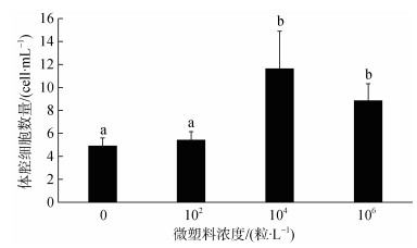 |
| 图 1 微塑料对黑海参体腔细胞数量的影响 Fig. 1 Effect of microplastics on the total coelomocyte count of Holothuria atra |
微塑料浓度对黑海参体腔细胞的吞噬活性有显著影响(P < 0.05, 图 2)。海水中微塑料浓度升高使黑海参体腔细胞的吞噬活性总体呈上升趋势。对照组黑海参体腔细胞的吞噬活性为(0.22±0.061) OD540/106cell, 水体中添加102、104粒/L微塑料时, 黑海参体腔细胞的吞噬活性虽高于对照组, 但变化不显著[(0.23± 0.065)、(0.22±0.064) OD540/106cell, P > 0.05], 当水体中微塑料浓度升至106粒/L时, 吞噬活性显著增加至(0.34±0.057) OD540/106cell(P < 0.05)。
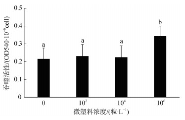 |
| 图 2 微塑料对黑海参体腔细胞吞噬活性影响 Fig. 2 Effect of microplastics on the phagocytic activity of coelomocyte in H. atra |
微塑料浓度对黑海参体腔液中酸性磷酸酶的活性有极显著影响(P=0.001, 图 3)。随着海水中微塑料添加浓度的增加, 黑海参体腔液中的酸性磷酸酶活性呈先增加后降低的趋势。未添加微塑料时, 黑海参体腔液中酸性磷酸酶的活性为(43.76±5.21) 金氏单位/gprot, 在微塑料浓度为102粒/L时, 酸性磷酸酶活性显著增加到(65.82±4.13) 金氏单位/gprot(P < 0.05), 当海水中添加104粒/L微塑料时, 酸性磷酸酶活性有所增加但变化不显著[(76.97 ± 18.46) 金氏单位/gprot, P > 0.05], 106粒/L时酸性磷酸酶活性显著降低但仍高于对照组[(59.12 ± 8.48) 金氏单位/gprot, P < 0.05]。
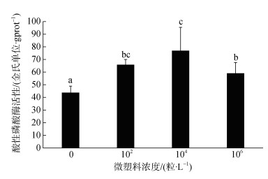 |
| 图 3 黑海参体腔液中酸性磷酸酶活性的变化 Fig. 3 Variation of ACP activity in coelomic fluid of H. atra |
如图 4所示, 微塑料浓度对黑海参体腔液中碱性磷酸酶活性的影响不显著(P > 0.05)。黑海参体腔液中碱性磷酸酶的活性随海水中微塑料浓度的增加呈上升趋势。对照组黑海参体腔液中碱性磷酸酶活性为(41.17±5.03) 金氏单位/gprot, 水体中添加102、104、106粒/L微塑料时, 碱性磷酸酶活性呈先上升后下降的趋势, 但未发生显著变化[(47.45± 11.76)、(35.42±5.94)和(37.10±7.78) 金氏单位/gprot, P > 0.05]。
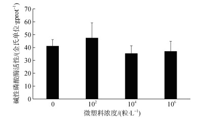 |
| 图 4 黑海参体腔液中碱性磷酸酶活性的变化 Fig. 4 Variation of AKP activity in coelomic fluid of H. atra |
微塑料浓度对黑海参体腔液中溶菌酶的活性有极显著影响(P < 0.001, 图 5)。随着海水中微塑料浓度的增加, 黑海参体腔液中溶菌酶活性总体呈先上升后下降的趋势。对照组黑海参体腔液中的溶菌酶活性为(96.97±5.12) U/mgprot, 海水中添加102粒/L微塑料时, 黑海参溶菌酶活性显著增加至(199.48±22.68) U/mgprot(P < 0.05), 微塑料浓度为104、106粒/L时, 溶菌酶活性有所降低但变化不显著[(174.56±16.89)、(176.47±14.02) U/mgprot, P > 0.05]。
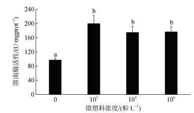 |
| 图 5 黑海参体腔液中溶菌酶活性变化 Fig. 5 Variation of LZM activity in coelomic fluid of H. atra |
水体中微塑料的浓度对黑海参体腔液中超氧化物歧化酶的活性有极显著影响(P < 0.001, 图 6)。海水中微塑料浓度升高使超氧化物歧化酶活性呈先增加后降低的趋势。对照组黑海参体腔液中超氧化物歧化酶活性为(29.05±0.74) U/mgprot, 海水中添加102粒/L微塑料时, 超氧化物歧化酶活性显著增加为(37.29±1.86) U/mgprot(P < 0.05), 当微塑料浓度升高为104粒/L时, 超氧化物歧化酶活性显著下降[(31.08±0.88) U/mgprot, P < 0.05], 但仍高于对照组, 海水中微塑料浓度为106粒/L时, 超氧化物歧化酶活性显著降低为(21.80±1.81) U/mgprot(P < 0.05), 且低于对照组。
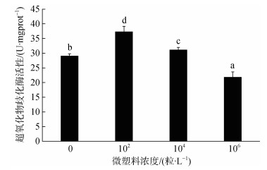 |
| 图 6 黑海参体腔液中超氧化物歧化酶活性的变化 Fig. 6 Variation of SOD activity in coelomic fluid of H. atra |
微塑料浓度对黑海参肠道淀粉酶活性有显著影响(P < 0.05, 图 7)。随着海水中微塑料浓度的增加, 黑海参肠道淀粉酶活性呈先增加后降低的趋势。对照组黑海参肠道淀粉酶的活性为(0.29±0.021) U/mgprot, 海水中添加102粒/L微塑料时, 淀粉酶活性未发生显著变化[(0.29±0.018) U/mgprot, P > 0.05], 在微塑料浓度为104粒/L时, 淀粉酶活性显著增加为(0.63±0.05) U/mgprot(P < 0.05), 当微塑料浓度升高为106粒/L时, 淀粉酶活性却显著降低为(0.15±0.49) U/mgprot(P < 0.05), 且低于对照组。
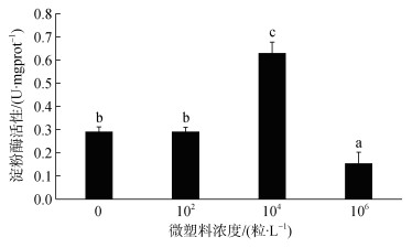 |
| 图 7 黑海参淀粉酶活性的变化 Fig. 7 Variation of AMS activity in the intestine of H. atra |
微塑料浓度对黑海参肠道胰蛋白酶活性影响不显著(P > 0.05, 图 8)。海水中微塑料浓度升高使黑海参肠道胰蛋白酶活性总体呈上升趋势。对照组肠道胰蛋白酶活性为(198.45±89.65) U/mgprot, 当海水中添加102、104粒/L微塑料时, 胰蛋白酶活性虽有所增加但均未发生显著变化(P > 0.05), 活性分别为(199.93±35.34)、(204.12±53.71) U/mgprot, 当海水中微塑料浓度增加为106粒/L时, 肠道胰蛋白酶活性显著升高为(319.23±96.35) U/mgprot(P < 0.05)。
 |
| 图 8 黑海参胰蛋白酶活性的变化 Fig. 8 Variation of TRY activity in the intestine of H. atra |
微塑料浓度对黑海参肠道脂肪酶活性的影响不显著(P=0.228, 图 9)。海水中微塑料浓度的升高使黑海参肠道脂肪酶活性总体呈下降趋势。对照组肠道脂肪酶活性为(1.76±0.32) U/gprot, 海水中添加102粒/L微塑料时, 脂肪酶活性降低至(1.26±0.31) U/ gprot, 但变化不显著(P > 0.05), 当微塑料浓度升高为104粒/L时, 脂肪酶活性为(1.31±0.28) U/gprot, 与102粒/L组脂肪酶活性相当(P > 0.05), 海水中微塑料浓度为106粒/L时, 脂肪酶活性最低且未发生显著变化[(1.17±0.46) U/gprot, P > 0.05]。
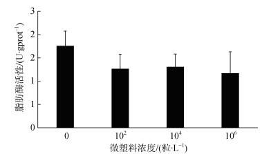 |
| 图 9 黑海参肠道脂肪酶活性变化 Fig. 9 Variation of LPS activity in the intestine of H. atra |
海水或沉积物中的微塑料可通过口或肛门进入海参体内, 并经由呼吸树进入体腔液中[15, 17]。海参体腔液中悬浮的多种体腔细胞和多种免疫因子构成海参的细胞免疫系统和体液免疫系统, 二者共同施行机体的免疫功能[25-26]。微塑料这种异物进入海参的体腔液后, 会激发海参的非特异性免疫应答, 产生一系列的免疫反应[17]。本研究将黑海参在不同浓度的聚苯乙烯暴露后, 测定了其体腔细胞数量、吞噬活性及体腔液中ACP、AKP、LZM和SOD的酶活性变化。
体腔细胞悬浮在海参的体腔液中, 其不仅具有吞噬、内陷和包埋入侵外源物质的作用, 而且能够分泌参与免疫反应的多种免疫因子和免疫酶, 是棘皮动物主要的非特异性防御指标[23, 27], 其数量和吞噬活性受环境因素的影响, 并在一定程度上反映机体的免疫力[28-31]。本研究中黑海参体腔细胞的数量在微塑料浓度升高至104、106粒/L时显著增加, 吞噬活性在微塑料浓度较高时也显著增加。该结果与贻贝(Mytilus edulis)在海水中接触铜之后血细胞吞噬活性增加的结果一致[32]。在虹鳟鱼中, 体内和体外的镉污染都会导致免疫细胞的吞噬活性的升高[33]。本实验中, 海水中添加的微塑料可通过呼吸和摄食作用进入黑海参体内, 黑海参增加体腔液中体腔细胞的数量, 并启动体腔细胞的吞噬作用, 产生大量的活性氧离子产物, 用于清除异物。
溶菌酶(LZM)是吞噬细胞杀菌的物质基础, 具有重要的免疫防御功能, 是衡量机体非特异性免疫的重要指标, 其活性常受到温度、pH和重金属离子等多种因素的影响[34-35]。当温度升高时, 大西洋比目鱼的LZM水平随之升高[36]; 低温骤变处理下, 凡纳滨对虾(Litopenaeus vannamei)的LZM活性显著低于对照组水平, 且温度越低, LSZ活力越低; 缺氧胁迫也会影响仿刺参呼吸树中LZM的活性[35]。在本研究中, 不同浓度的微塑料暴露均使黑海参体腔液中LZM活性显著升高, Mohsen等研究表明当纤维微塑料从水体中转移至仿刺参体内时, 仿刺参体腔液中的LZM活性也高于对照组[17]; 暴露于纳米聚苯乙烯微球中的双壳贻贝(Mytilus galloprovincialis)也具有较高的LZM活性[37]。
酸性磷酸酶(ACP)是巨噬细胞内溶酶体的标志酶, 可协助降解外源物质[38], 并可增强细胞对异物的识别, 加快吞噬细胞对异物的吞噬和降解速度[39]。碱性磷酸酶(AKP)可催化磷酸单酯的水解反应及磷酸基团的转移反应, 且对底物专一性要求较低, 是重要的解毒系统, 与一些营养物质的吸收转运有关[30, 40]。微塑料浓度的增加对黑海参体腔液的ACP、AKP活性均产生了影响, 而仿刺参体腔液中ACP及AKP的活性在聚酯纤维微塑料(25、40粒/mL)暴露后未发生显著变化[17]。此外, 已有研究发现七彩神仙鱼和中华绒螯蟹的ACP和AKP活性在微塑料暴露后显著增加[41-42]。因此, 微塑料对生物体内ACP和AKP活性的影响与物种等因素有关。ACP和AKP活性的增加表明黑海参机体内对外源物质的第一道防御增强, 代谢强度提高, 需要更多的能量供应。黑海参在代谢过程中需要消耗更多的ATP来适应外部环境中微塑料对其呼吸系统的干扰, 而ATP合成所需的磷酸可以通过ACP和AKP水解磷酸单酯生成[43]。此外, ACP、AKP活性随海水中微塑料浓度的增加呈先增强后减弱的趋势, 这与白斑综合征病毒(WSSV)感染日本囊对虾ACP活性变化一致[44]。磷酸酶主要位于膜系统上, 中华绒螯蟹感染白斑综合征病毒(WSSV)时间延长可导致溶酶体膜结构受损, 从而使ACP、AKP酶活性显著降低[40]。黑海参体腔液中ACP、AKP活性分别在水体中微塑料增加至104、106粒/L后降低, 可能是微塑料在黑海参体内含量的增加损伤了体腔细胞的细胞结构, 导致溶酶体膜受损, 渗透性改变, 进而使ACP、AKP活性受到限制[44]。
超氧化物歧化酶(SOD)作为一种重要的抗氧化酶, 主要参与清除动物体内过多的活性氧自由基, 保持细胞免受损害, 使细胞能正常合成各种酶类, 对增强吞噬细胞活性和整个机体的免疫功能具有重要的作用[45], 病原微生物和多种环境胁迫因子均会引发机体SOD活性的增加[28, 40, 43-44]。微塑料也会诱导水生生物产生氧化应激反应, 如斑马鱼(Danio rerio)在微塑料暴露后, 体内SOD活性显著增加[46]。在本研究中, 随着微塑料浓度的增加, 黑海参体腔液中SOD的活性呈现先升高后降低的变化, 这与丛聪研究溶壁微球菌和假交替单胞菌对仿刺参体腔液中SOD活性影响的结果一致[47]。此外, 海马(Hippocampus kuda Bleeker)、罗非鱼(Oreochromis niloticus)体内的SOD活性随微塑料暴露时间的增加也呈先升高后降低的趋势[48-49]。水体中微塑料为102、104粒/L时, SOD活性显著高于对照组, 说明微塑料引起了黑海参SOD系统的应激反应, SOD活性增强可以在一定程度上清除黑海参体内在微塑料暴露后产生的活性氧自由基, 是海参对外部环境变化产生的适应对策。而SOD活性在微塑料浓度为106粒/L时显著低于对照组及102、104粒/L实验组, 这与高浓度微塑料使海参体内吞噬活性、呼吸爆发活性增强而持续产生的大量活性氧自由基得不到及时有效地清除有关, 活性氧自由基对机体造成了氧化损伤, 削弱了黑海参SOD的免疫应答能力[48-49]。
3.2 微塑料对黑海参消化酶活性的影响消化酶的活性受多种因素的影响, 是衡量动物摄食能力、营养和生理状态常用的指标之一, 其高低与营养吸收、生长和发育密切相关, 可用来判断动物对环境的适应情况[50-51]。动物可以通过调节自身的酶活性以提高从周围环境中获取营养成分的能力[52]。沉积性食性的海参依靠触手主动摄食沉积物中的无机物(硅或钙)、单细胞藻类、动植物有机碎屑、原生动物、细菌和腐殖质等[49]。在天然饵料或人工饲料中, 对海参生长贡献最大的是蛋白质, 其次是碳水化合物、脂肪等物质, 相关研究也多集中在蛋白酶、淀粉酶和脂肪酶等[53]。
海参的消化酶活性与温度、盐度、肠道菌群和摄食量等多种因素有关[35, 41, 54]。微塑料摄入会使生物产生饱腹感[37], 改变其体内消化酶的活性[55], 进而影响生物对营养物质的消化和吸收。Romano等发现接触聚氯乙烯(PVC)微塑料(0.5、1.0 mg/L)的爪哇四须
本文从免疫、消化等方面探讨了水体中添加聚苯乙烯微塑料对黑海参生理的影响, 微塑料浓度对黑海参的体腔细胞数量、吞噬活性、ACP、LZM和SOD活性均有显著影响, 表明微塑料暴露会引发黑海参的免疫应答; 但随着微塑料浓度的增加, 黑海参的体腔细胞数量、ACP、LZM和SOD活性均呈现先升高后降低的趋势, 尤其是当微塑料浓度达到106粒/L时, SOD活性降到了显著低于对照组的水平, 这表明黑海参的免疫防御反应对微塑料暴露有浓度效应, 且过高的微塑料浓度引发的免疫应答可能对机体造成了氧化损伤。消化生理指标的变化表明, 在微塑料暴露条件下, 黑海参可能会选择易于消化的营养物质, 例如淀粉和蛋白质, 而不是脂质, 以便快速获取能量来适应环境中微塑料的存在。
| [1] |
SEBILLE E V, WILCOX C, LEBRETON L, et al. A global inventory of small floating plastic debris[J]. Environmental Research Letters, 2015, 10(12): 124006. DOI:10.1088/1748-9326/10/12/124006 |
| [2] |
KNUTSEN H, CYVIN J B, TOTLAND C, et al. Microplastic accumulation by tube-dwelling, suspension feeding polychaetes from the sediment surface: A case study from the Norwegian Continental Shelf[J]. Marine Environmental Research, 2020, 161: 105073. DOI:10.1016/j.marenvres.2020.105073 |
| [3] |
TATA T, BELABED B E, BOUOUDINA M, et al. Occurrence and characterization of surface sediment microplastics and litter from North African coasts of Mediterranean Sea: Preliminary research and first evidence[J]. Science of the Total Environment, 2020, 713: 136664. DOI:10.1016/j.scitotenv.2020.136664 |
| [4] |
COSTA J P D, SANTOS P S M, DUARTE A C, et al. (Nano)plastics in the environment-Sources, fates and effects[J]. Science of the Total Environment, 2016, 566-567: 15-26. DOI:10.1016/j.scitotenv.2016.05.041 |
| [5] |
TANG Y Q, LIU Y G, CHEN Y, et al. A review: Research progress on microplastic pollutants in aquatic environments[J]. Science of the Total Environment, 2020, 142572. |
| [6] |
BOUCHER J, FRIOT D. Primary Microplastics in the Oceans: A Global Evaluation of Sources[M]. Gland, Switzerland: International Union for Conservation of Nature, 2017: 227-229.
|
| [7] |
SJOLLEMA S B, REDONDO-HASSELERHARM P, LESLIE H A, et al. Do plastic particles affect microalgal photosynthesis and growth?[J]. Aquatic Toxicology, 2016, 170: 259-261. DOI:10.1016/j.aquatox.2015.12.002 |
| [8] |
CARUSO G. Microplastics as vectors of contaminants[J]. Marine Pollution Bulletin, 2019, 146(7): 921-924. |
| [9] |
VON M N, BURKHARDT-HOLM P, KÖHLER A. Uptake and effects of microplastics on cells and tissue of the blue mussel Mytilus edulis L. after an experimental exposure[J]. Environmental Science & Technology, 2012, 46(20): 11327-11335. |
| [10] |
FACKELMANN G, SOMMER S. Microplastics and the gut microbiome: How chronically exposed species may suffer from gut dysbiosis[J]. Marine Pollution Bulletin, 2019, 143: 193-203. DOI:10.1016/j.marpolbul.2019.04.030 |
| [11] |
YIN L, CHEN B, XIA B, et al. Polystyrene microplastics alter the behavior, energy reserve and nutritional composition of marine jacopever (Sebastes schlegelii)[J]. Journal of Hazardous Materials, 2018, 360: 97-105. DOI:10.1016/j.jhazmat.2018.07.110 |
| [12] |
JABEEN K, LI B, CHEN Q, et al. Effects of virgin microplastics on goldfish (Carassius auratus)[J]. Chemosphere, 2018, 213: 323-332. DOI:10.1016/j.chemosphere.2018.09.031 |
| [13] |
SUN J H, XIA S D, NING Y, et al. Effects of microplastics and attached heavy metals on growth, immunity, and heavy metal accumulation in the yellow seahorse, Hippocampus kuda[J]. Marine Pollution Bulletin, 2019, 149: 110510. DOI:10.1016/j.marpolbul.2019.110510 |
| [14] |
GARDON T, REISSER C, SOYEZ C, et al. Microplastics affect energy balance and gametogenesis in the pearl oyster Pinctada margaritifera[J]. Environmental Science & Technology, 2018, 52(9): 5277-5286. |
| [15] |
IWALAYE O A, MOODLEY G K, ROBERTSON-ANDERSSON D V. The possible routes of microplastics uptake in sea cucumber Holothuria cinerascens (Brandt,1835)[J]. Environmental Pollution, 2020, 264: 114644. DOI:10.1016/j.envpol.2020.114644 |
| [16] |
MOHSEN M, WANG Q, ZHANG L, et al. Microplastic ingestion by the farmed sea cucumber Apostichopus japonicus in China[J]. Environmental Pollution, 2018, 245: 1071-1078. |
| [17] |
MOHSEN M, ZHANG L, SUN L, et al. Microplastic fibers transfer from the water to the internal fluid of the sea cucumber Apostichopus japonicus[J]. Environmental Pollution, 2020, 257: 113606. DOI:10.1016/j.envpol.2019.113606 |
| [18] |
廖玉麟. 中国动物志[M]. 北京: 科学出版社, 1997: 115-117. LIAO Yulin. The Faunal Monographs of China[M]. Beijing: Science Press, 1997: 115-117. |
| [19] |
PURCELL S W, SAMYN Y, CONAND C. Commercially Important Sea Cucumbers of the World[M]. Rome, Italy: Food and Agriculture Organization, 2012: 128-140.
|
| [20] |
PURCELL S W, CONAND C, UTHICKE S, et al. Ecological Roles of Exploited Sea Cucumbers[M]. Oceanography and Marine Biology, 2016, 54: 367-386.
|
| [21] |
TORAL-GRANDA V, LOVATELLI A, VASCONCELLOS M, et al. Sea Cucumbers: A Global Review on Fishery and Trade[M]. Rome, Italy: FAO Fisheries and Aquaculture Technical Paper, 2008: 7-118.
|
| [22] |
ESMAT A Y, SAID M M, SOLIMAN A A, et al. Bioactive compounds, antioxidant potential, and hepatoprotective activity of sea cucumber (Holothuria atra) against thioacetamide intoxication in rats[J]. Nutrition, 2013, 29(1): 258-267. DOI:10.1016/j.nut.2012.06.004 |
| [23] |
谭杰. 刺参(Apostichopus japonicus)苗种繁育关键技术及幼参对操作胁迫的免疫应答研究[D]. 青岛: 中国海洋大学, 2015. TAN Jie. Study on the critical technology of Apostichopus japonicus artificial breeding and immune response of juvenile to handling stress[D]. Qingdao: Ocean University of China, 2015. |
| [24] |
ZHAO Y, ZHANG W, XU W, et al. Effects of potential probiotic Bacillus subtilis T13 on growth, immunity and disease resistance against Vibrio splendidus infection in juvenile sea cucumber Apostichopus japonicus[J]. Fish & Shellfish Immunology, 2012, 32(5): 750-755. |
| [25] |
叶海斌, 樊英, 李天保, 等. 刺参免疫防御机制研究进展[J]. 安徽农业科学, 2018, 46(16): 27-29. YE Haibin, FAN Ying, LI Tianbao, et al. Study progress on immunity defense mechanism of Apostichopus japonicus[J]. Journal of Anhui Agricultural Sciences, 2018, 46(16): 27-29. DOI:10.3969/j.issn.0517-6611.2018.16.009 |
| [26] |
ELLIS R P, PARRY H, SPICER J I, et al. Immunological function in marine invertebrates: Responses to environmental perturbation[J]. Fish & Shellfish Immunology, 2011, 30: 1209-1222. |
| [27] |
DOLMATOVA L S, ELISEYKINA M G, TIMCHENKO N F, et al. Generation of reactive oxygen species in different fractions of the coelomocytes of holothurian Eupentacta fraudatrix in response to the thermostable toxin of Yersinia pseudotuberculosis in vitro[J]. Chinese Journal of Oceanology and Limnology, 2003, 21: 293-304. DOI:10.1007/BF02860423 |
| [28] |
WANG F, YANG H, GAO F, et al. Effects of acute temperature or salinity stress on the immune response in sea cucumber, Apostichopus japonicus[J]. Comparative Biochemistry and Physiology Part A: Molecular & Integrative Physiology, 2008, 151(4): 491-498. |
| [29] |
李华, 陈静, 陆佳, 等. 仿刺参体腔细胞和血细胞类型及体腔细胞数量研究[J]. 水生生物学报, 2009, 33(2): 208-213. LI Hua, CHEN Jing, LU Jia, et al. Type and quantity of blood cells and coelomocytes in Apostichopus japonicus[J]. Acta Hydrobiologica Sinica, 2009, 33(2): 208-213. |
| [30] |
贾晨晨. 菊芋全粉对仿刺参生长、免疫力及肠道微生物的影响[D]. 青岛: 中国科学院大学(中国科学院烟台海岸带研究所), 2019. JIA Chenchen. Effects of Jerusalem artichoke powder on the growth performance, immune responses and gastrointestinal microbiota of sea cucumber Apostichopus japonicus[D]. Qingdao: University of Chinese Academy of Sciences (Yantai Institute of Costal Zone Research), 2019. |
| [31] |
田景玉, 丁月, 张晶, 等. 紫外线照射对刺参体腔细胞吞噬活性的影响[J]. 大连工业大学学报, 2017, 36(6): 406-410. TIAN Jingyu, DING Yue, ZHANG Jing, et al. Effect of UVA on the phagocytic activity of coelomocyte in sea cucumber Stichopus japonicus[J]. Journal of Dalian Polytechnic University, 2017, 36(6): 406-410. DOI:10.3969/j.issn.1674-1404.2017.06.005 |
| [32] |
PIPE R K, COLES J A, CARISSAN F M M, et al. Copper induced immunomodulation in the marine mussel, Mytilus edulis[J]. Aquatic Toxicology, 1999, 46(1): 43-54. DOI:10.1016/S0166-445X(98)00114-3 |
| [33] |
ZELIKOFF J T, BOWSER D, SQUIBB K S, et al. Immunotoxicity of low-level cadmium exposure in fish: An alternative animal model for immunotoxicological studies[J]. Journal of Toxicology and Environmental Health, Part A Current Issues, 1995, 45(3): 235-248. DOI:10.1080/15287399509531993 |
| [34] |
MÖCK A, PETERS G. Lysozyme activity in rainbow trout, Oncorhynchus mykiss (Walbaum), stressed by handling, transport and water pollution[J]. Journal of Fish Biology, 1990, 37(6): 873-885. DOI:10.1111/j.1095-8649.1990.tb03591.x |
| [35] |
HUO D, SUN L, RU X, et al. Impact of hypoxia stress on the physiological responses of sea cucumber Apostichopus japonicus: Respiration, digestion, immunity and oxidative damage[J]. PeerJ, 2018, 6: 4651. DOI:10.7717/peerj.4651 |
| [36] |
LANGSTON A L, HOARE R, STEFANSSON M, et al. The effect of temperature on non-specific defence parameters of three strains of juvenile Atlantic halibut (Hippoglossus hippoglossus L.)[J]. Fish & Shellfish Immunology, 2002, 12(1): 61-76. |
| [37] |
CANESI L, CIACCI C, BERGAMI E, et al. Evidence for immunomodulation and apoptotic processes induced by cationic polystyrene nanoparticles in the hemocytes of the marine bivalve Mytilus[J]. Marine Environmental Research, 2015, 111: 34-40. DOI:10.1016/j.marenvres.2015.06.008 |
| [38] |
MENG Z, SHAO J, XIANG L. CpG oligodeoxynucleotides activate grass carp (Ctenopharyngodon idellus) macrophages[J]. Developmental and Comparative Immunology, 2003, 27(4): 313-321. DOI:10.1016/S0145-305X(02)00104-0 |
| [39] |
李海兵, 宋晓玲, 韦篙, 等. WSSV感染对克氏原鳌虾血细胞吞噬和肝胰腺磷酸酶活性的影响[J]. 渔业科学进展, 2011, 32(2): 78-82. LI Haibing, SONG Xiaoling, WEI Gao, et al. Impacts of WSSV on the activity of haemocyte phagocytosis and hepatopancreas phosphatase in Procambarus clarkia[J]. Progress in Fishery Sciences, 2011, 32(2): 78-82. DOI:10.3969/j.issn.1000-7075.2011.02.013 |
| [40] |
孙婷. 白斑综合征病毒对中华绒螯蟹的免疫毒性作用[D]. 上海: 华东师范大学, 2013. SUN Ting. Effects of WSSV on immunotoxicity of Eriocheir sinensis[D]. Shanghai: East China Normal University, 2013. |
| [41] |
WEN B, ZHANG N, JIN S R, et al. Microplastics have a more profound impact than elevated temperatures on the predatory performance, digestion and energy metabolism of an Amazonian cichlid[J]. Aquatic Toxicology, 2018, 195: 67-76. DOI:10.1016/j.aquatox.2017.12.010 |
| [42] |
LIU Z, YU P, CAI M, et al. Effects of microplastics on the innate immunity and intestinal microflora of juvenile Eriocheir sinensis[J]. Science of the Total Environment, 2019, 685: 836-846. DOI:10.1016/j.scitotenv.2019.06.265 |
| [43] |
郑慧, 李彬, 荣小军, 等. 盐度和溶解氧对刺参非特异性免疫酶活性的影响[J]. 渔业科学进展, 2014, 35(1): 118-124. ZHENG Hui, LI Bin, RONG Xiaojun, et al. Effects of salinity and dissolved oxygen variation on the non-specific immune response of Apostichopus japonicus[J]. Progress in Fishery Sciences, 2014, 35(1): 118-124. DOI:10.3969/j.issn.1000-7075.2014.01.017 |
| [44] |
张曼, 宋维彦, 王军. 白斑综合征病毒对日本囊对虾仔虾免疫相关酶活性的影响[J]. 厦门大学学报(自然科学版), 2011, 50(1): 117-122. ZHANG Man, SONG Weiyan, WANG Jun. Effects of WSSV on enzymes activities related with immunity of Marsupenaeus japonicus postlarvae[J]. Journal of Xiamen University (Natural Science), 2011, 50(1): 117-122. |
| [45] |
董晓亮, 李成林, 赵斌, 等. 低盐胁迫对刺参非特异性免疫酶活性及抗菌活力的影响[J]. 渔业科学进展, 2013, 34(3): 82-87. DONG Xiaoliang, LI Chenglin, ZHAO Bin, et al. Effects of low salinity stress on the activities of non-specific immune enzymes and antibacterial activity of sea cucumber Apostichopus japonicus[J]. Progress in Fishery Sciences, 2013, 34(3): 82-87. DOI:10.3969/j.issn.1000-7075.2013.03.011 |
| [46] |
LU Y, ZHANG Y, DENG Y, et al. Uptake and accumulation of polystyrene microplastics in zebrafish (Danio rerio) and toxic effects in liver[J]. Environmental Science & Technology, 2016, 50(7): 4054-4060. |
| [47] |
丛聪. 仿刺参(Apostichopus japonicus)体腔液的抗菌特性[D]. 大连: 大连海洋大学, 2015. CONG Cong. Antibacterial analysis of coelomic fluid from the sea cucumber (Apostichopus japonicus)[D]. Dalian: Dalian Ocean University, 2015. |
| [48] |
SUN J H, XIA S D, NING Y, et al. Effects of microplastics and attached heavy metals on growth, immunity, and heavy metal accumulation in the yellow seahorse, Hippocampus kuda Bleeker[J]. Marine Pollution Bulletin, 2019, 149: 110510. DOI:10.1016/j.marpolbul.2019.110510 |
| [49] |
DING J, ZHANG S, RAZANAJATOVO R M, et al. Accumulation, tissue distribution, and biochemical effects of polystyrene microplastics in the freshwater fish red tilapia (Oreochromis niloticus)[J]. Environmental Pollution, 2018, 238: 1-9. DOI:10.1016/j.envpol.2018.03.001 |
| [50] |
FU X Y, XUE C H, MIAO B C, et al. Characterization of proteases from the digestive tract of sea cucumber (Stichopus japonicus): High alkaline protease activity[J]. Aquaculture, 2005, 246(1/4): 321-329. |
| [51] |
杨心愿, 林承刚, 宋肖跃, 等. 不同产地浒苔对幼刺参生长、消化和非特异性免疫的影响[J]. 海洋与湖沼, 2018, 49(5): 1103-1108. YANG Xinyuan, LIN Chenggang, SONG Xiaoyue, et al. Effects of Ulva Prolifera from different sites on growth, digestion and non-specific immunity of juvenile sea cucumber (Apostichopus japonicus)[J]. Oceanologia et Limnologia Sinica, 2018, 49(5): 1103-1108. |
| [52] |
HASSETT R P, LANDRY M R. Seasonal changes in feeding rate, digestive enzyme activity, and assimilation efficiency of Calanus pacificus[J]. Marine Ecology Progress Series, 1990, 62(3): 203-210. |
| [53] |
包鹏云. 刺参常用饲料原料营养价值评价及其对主要营养物质有效利用的研究[D]. 大连: 大连理工大学, 2018. BAO Pengyun. Studies on the nutritive value of commonly used feed ingredients for sea cucumber (Apostichopus japonicus) and the efficient use of major nutrients in the feeds[D]. Dalian: Dalian University of Technology, 2018. |
| [54] |
GAO F, YANG H S, XU Q, et al. Effect of water temperature on digestive enzyme activity and gut mass in sea cucumber Apostichopus japonicus (Selenka), with special reference to aestivation[J]. Chinese Journal of Oceanology & Limnology, 2009, 4: 714-722. |
| [55] |
ROMANO N, ASHIKIN M, TEH J C, et al. Effects of pristine polyvinyl chloride fragments on whole body histology and protease activity in silver barb Barbodes gonionotus fry[J]. Environmental Pollution, 2017, 237: 1106-1111. |
| [56] |
LI E, CHEN L, ZENG C, et al. Comparison of digestive and antioxidant enzymes activities, haemolymph oxyhemocyanin contents and hepatopancreas histology of white shrimp, Litopenaeus vannamei, at various salinities[J]. Aquaculture, 2008, 274(1): 80-86. DOI:10.1016/j.aquaculture.2007.11.001 |
| [57] |
MATTSON M P. Hormesis defined[J]. Ageing Research Reviews, 2008, 7(1): 1-7. DOI:10.1016/j.arr.2007.08.007 |
 2021, Vol. 45


